 Jovian Labs
SEM/TEM particle size analysis with EU nanomaterial screening
Turn electron microscope images into a particle size distribution report in about ten seconds per image: D10, D50 and D90 with 95% confidence intervals, minimum and maximum Feret diameters, shape factors, nine automatic quality checks and a screening against the EU definition of nanomaterial.
Run it on Apify Example report (PDF)
EU nanomaterial screening
Under Commission Recommendation 2022/C 229/01, a material meets criterion (a) when 50% or more of its particles, counted by number, have at least one external dimension between 1 and 100 nm. Each run reports the share of particles whose minimum Feret diameter is 100 nm or less, a Wilson confidence interval, a wider interval that adds calibration error and edge blur, the median minimum Feret, and a verdict: meets, not met, or borderline when the interval includes 50%. Read the guide.
What you get
| Output | Content |
|---|---|
| PDF report | Results and confidence intervals, nanomaterial screening, distribution and cumulative curve, results per image, quality checks, method, traceability |
| Per-particle CSV | Status, equivalent diameter, min/max Feret, aspect ratio, circularity, solidity, position |
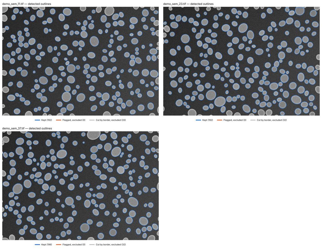
| Control sheet | Every image with the detected outlines |
| JSON and dataset row | All statistics in machine-readable form, for scripts, spreadsheets and AI agents |
Accuracy
- Synthetic SEM images with known sizes (the free demo): 580 particles measured for 578 present, D50 within +3.7% of the truth.
- Nanomaterial screening on synthetic samples with a known share at or below 100 nm (96%, 70%, 62%, 10%): measured within 1 to 6 points, always inside the reported interval; the 62% sample is correctly reported as borderline.
- Limit: strongly agglomerated powders read high, and the agglomerate check turns amber or red.
Control sheet from the demo
Questions
Which images can I use?
SEM or TEM images of one sample, all at the same magnification, as TIFF, PNG or JPG. Original Zeiss or Thermo Fisher TIFF files are calibrated from their metadata; otherwise give the scale bar length or the pixel size.
How many particles should I measure?
At least 300 for a size distribution and 500 for the nanomaterial screening. The report shows the precision reached on D50 and how many particles a ±2 % precision would need.
Is the nanomaterial verdict a regulatory determination?
No. It screens criterion (a) of the EU definition on the particles measured, with its uncertainty. It does not assess the volume specific surface area, dimensions below 1 nm or the thickness of platelets lying flat.
What does it cost?
1 US dollar per image analysed, billed by Apify. Demo runs on synthetic images are free.
Open the Actor on Apify and press Start with an empty image list: it analyses three synthetic images and compares the result with the truth.